
Technology Overview
technology
Overview
Since the early 1990s, there has been a growing recognition that optical interferometry can be applied to medicine for cross-sectional, or tomographic, imaging. The basis for this imaging method is that some near infrared (NIR) wavelengths can penetrate living tissues for very substantial distances. These wavelengths lie within the so-called "therapeutic window," which also allows for some light-based therapies.
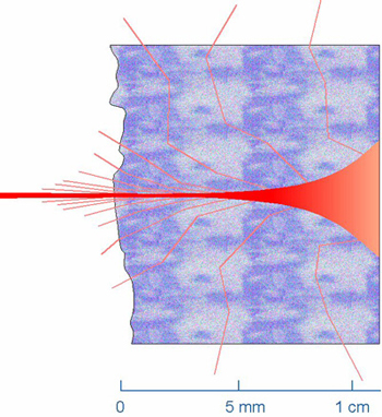
A focused NIR light beam entering a tissue remains concentrated for its first few millimeters of propagation before scattering causes the light to spread significantly. The depth of penetration is the greatest for wavelengths that lie in the “therapeutic window.”
In light-based tomography, optical interferometry plays the role of optical ranging. Although optical interferometry had long been utilized in non-medical fields and known as Optical Coherence Domain Reflectometry (OCDR), not until the 1990s did we see its first medical application in ophthalmology, a fruition of the pioneering work by J. G. Fujimoto et al. This modality has come to be known as Optical Coherence Tomography (OCT) in the arena of medical imaging.
An analogy can be drawn between OCT and ultrasound imaging in which the information carrier is sound waves instead of light. As the wavelength of the light is at least three orders of magnitude shorter than that of the sound wave, the imaging resolution of OCT is much higher, on the order of microns, making it ideal for the visualization of microanatomy and ultimately even cellular structures.
In order to realize the potential of OCT in a broader medical field some technical hurdles remain to be overcome. The need for better image clarity, higher resolution and greater imaging depth has continued to push the boundaries of fiber optics, signal processing and computing algorithms. For endoscopic applications, the miniaturization of the optical probe poses an additional technical challenge.
Tomophase OCT Technology
Tomophase OCT imaging System, or, OCTIS™, is a proprietary technology platform based on a series of innovations in fiber-optics, signal processing, computing algorithm and optical probe design. These innovations aim at providing more advanced imaging capabilities while avoiding the shortcomings of existing OCT technology.
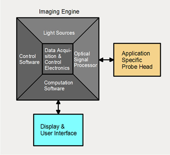
Tomophase OCTIS is composed of an imaging engine which is common to many applications, a user interface and an application specific optical probe or catheter.
The advantages and uniqueness of Tomophase technology stem, in part, from a proprietary optical imaging engine with an optical cross-correlator that circumvents the conventional optical interferometer. The Tomophase imagine engine possesses unique self-cancellation mechanism for the rejection of common-mode noise generated by the light sources as well as the movement of the optical fibers. As a result, our imaging engine provides superior optical signal quality compared to the conventional technology.
This optical imaging engine, coupled with our proprietary signal processing algorithm, allows us to retrieve not only the first-order tomogram, as with the conventional OCT technology, but also high-order imageries including phase-resolved tomography and differential spectral-absorbance mapping in imaged cross-sections.
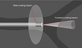
The proprietary Tomophase imaging probe provides the flexibility of scanning the light beam in either a forward-looking or a sideward-looking direction. Linked with the imaging engine, one can acquire tomographic tissue images of tissues located in both areas.
A Tomophase OCT Imaging system is complete with the addition of an application-specific catheter and a display device. A Tomophase proprietary design allows for the miniaturization of a highly flexible light delivery and collection catheter for imaging inside airways through the use of bronchoscopes. This miniature imaging catheter can direct the NIR beam to scan in either a forward-looking or a sideward-looking direction. These two scan directions are switchable through lightwave control at the proximal end of the catheter system. This allows the user to easily assess tissues located in the front and to the side of the catheter with a single procedure.